
Condition-Specific Pain Management
What is Right Knee Anatomy? Key Components and Importance Explained
Introduction
If you’re struggling with knee pain, you’re not alone; understanding the anatomy of your knee can be the first step toward relief. This joint, made up of bones, ligaments, tendons, and cartilage, is crucial for everyday activities like walking and running, and we understand how frustrating it can be when it hurts. As you face the challenges of knee discomfort, you might wonder how understanding your knee’s anatomy can help you find the right treatment and improve your recovery. By exploring these key components, we can uncover ways to manage your pain effectively and help you regain your quality of life.
Define Right Knee Anatomy
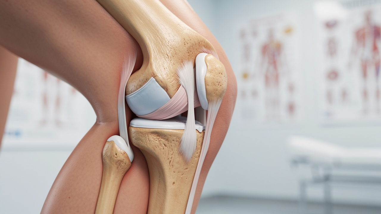
If you’re struggling with knee pain, understanding the right knee anatomy can seem overwhelming, but it’s a crucial step toward finding relief. The knee joint, which includes the patella, femur, and tibia, is one of the largest and most complex articulations in the human body. These bones work together, supported by ligaments, tendons, and cartilage, to facilitate movement and provide stability. This joint functions as a hinge, allowing for flexion and extension while also permitting a limited range of rotation.
We know that each person’s experience with knee pain is unique, and understanding the right knee anatomy is essential for addressing your pain and finding the appropriate treatment. Recent advancements in joint anatomy research are helping us tailor treatments just for you, especially in the context of joint replacement surgeries. For instance, the creation of custom 3D-printed implants designed for individual joint anatomy has shown promise in improving surgical results and patient satisfaction at Ponte Vedra Spine & Pain Center.
Case studies illustrate the effectiveness of these innovations; patients receiving personalized implants often report improved functionality and reduced recovery times. A thorough understanding of joint anatomy not only aids in surgical precision but also enhances rehabilitation strategies, ultimately leading to better patient outcomes. At Ponte Vedra Spine & Pain Center, we’re committed to using the latest technologies and personalized treatment plans to help you overcome knee-related challenges. With these advancements, you can feel hopeful about your journey to recovery and improved quality of life.
Explore Key Components of the Right Knee
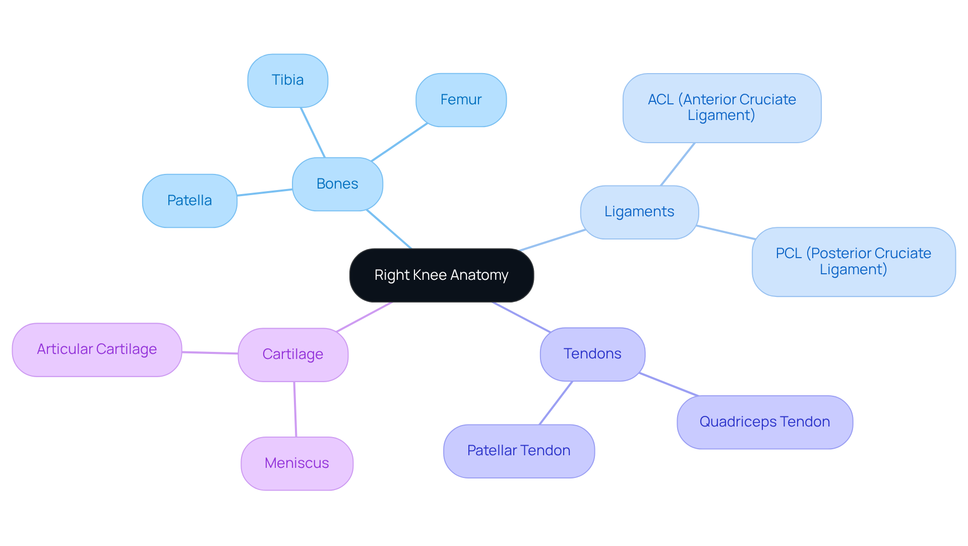
If you’re feeling discomfort in your knee, you’re not alone; many people face similar challenges that can impact daily life. The right knee anatomy is a complex joint composed of several key components, each essential for its proper function.
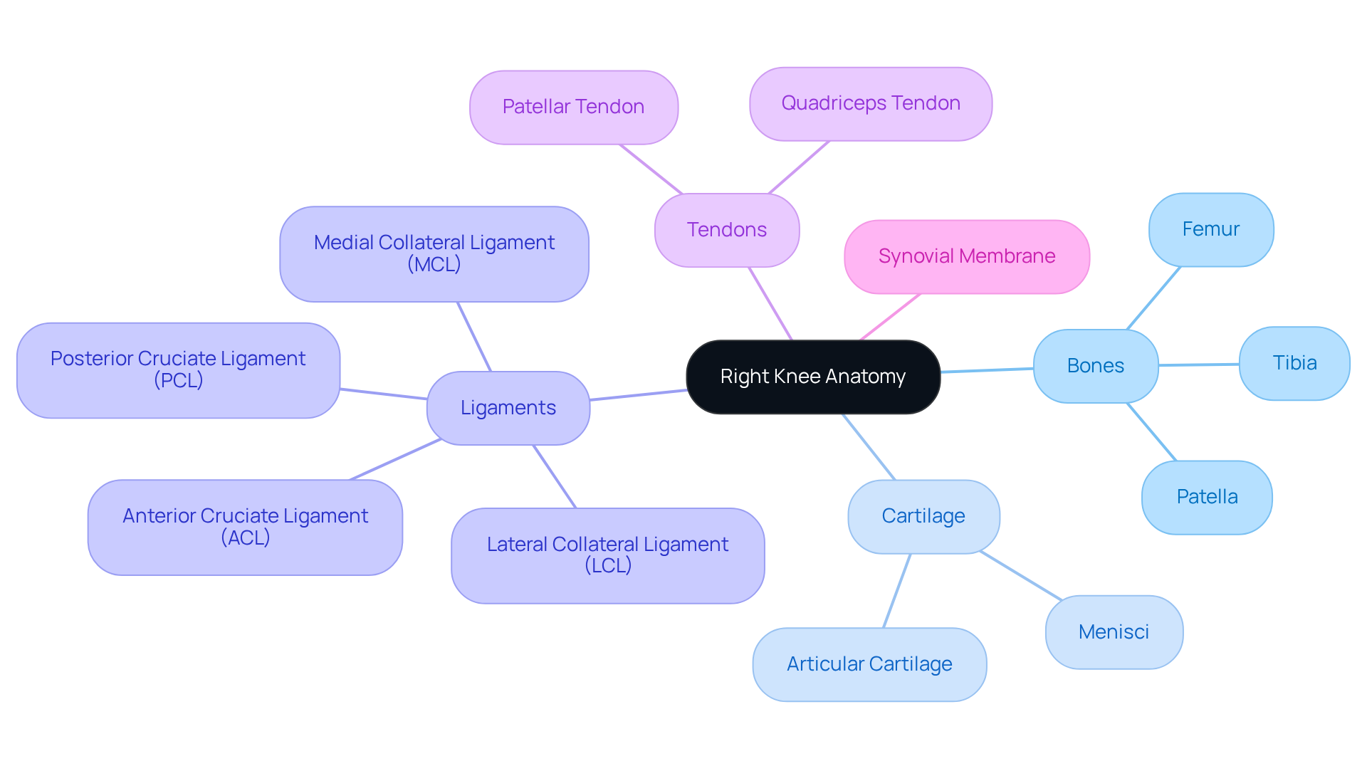
The structural framework of the joint consists of the following:
- Femur: The upper bone that connects to the hip.
- Tibia: The lower bone that connects to the ankle.
- Patella: The kneecap that sits anteriorly, providing protection and aiding in movement.
Articular cartilage covers the ends of the femur and tibia, offering a smooth surface for movement and acting as a shock absorber. Additionally, the menisci-two crescent-shaped cartilage structures-further cushion the joint and stabilize it during weight-bearing activities. Many people struggle with the pain and limitations caused by osteoarthritis, feeling overwhelmed and unsure of how to find relief. Understanding the components of the right knee anatomy can feel daunting, but it’s a crucial step in addressing the pain and limitations you may be experiencing.
Stability in the joint is provided by four primary ligaments:
- Anterior cruciate ligament (ACL)
- Posterior cruciate ligament (PCL)
- Medial collateral ligament (MCL)
- Lateral collateral ligament (LCL)
These ligaments connect the femur to the tibia, ensuring stability during various movements.
Tendons serve as connectors between muscles and bones. The quadriceps tendon attaches the quadriceps muscle to the patella, while the patellar tendon connects the patella to the tibia. These tendons play a vital role in helping you move freely, allowing you to enjoy everyday activities without discomfort.
The synovial membrane lines the capsule surrounding the articulation and produces synovial fluid, which lubricates the area, reducing friction during movement. This lubrication is essential for maintaining joint health and function.
Fortunately, there are innovative treatments available that can help restore comfort and improve your quality of life. Recent studies suggest that Genicular Artery Embolization (GAE) is a promising minimally invasive technique that can significantly alleviate discomfort for individuals suffering from arthritis in the joint. If you experience severe or persistent pain in your joints, it is essential to seek medical attention to determine the underlying cause and explore comprehensive management solutions available at Ponte Vedra Spine & Pain Center. Taking the first step towards understanding your knee health can lead to a brighter, more active future.
Understand the Functional Importance of Right Knee Anatomy
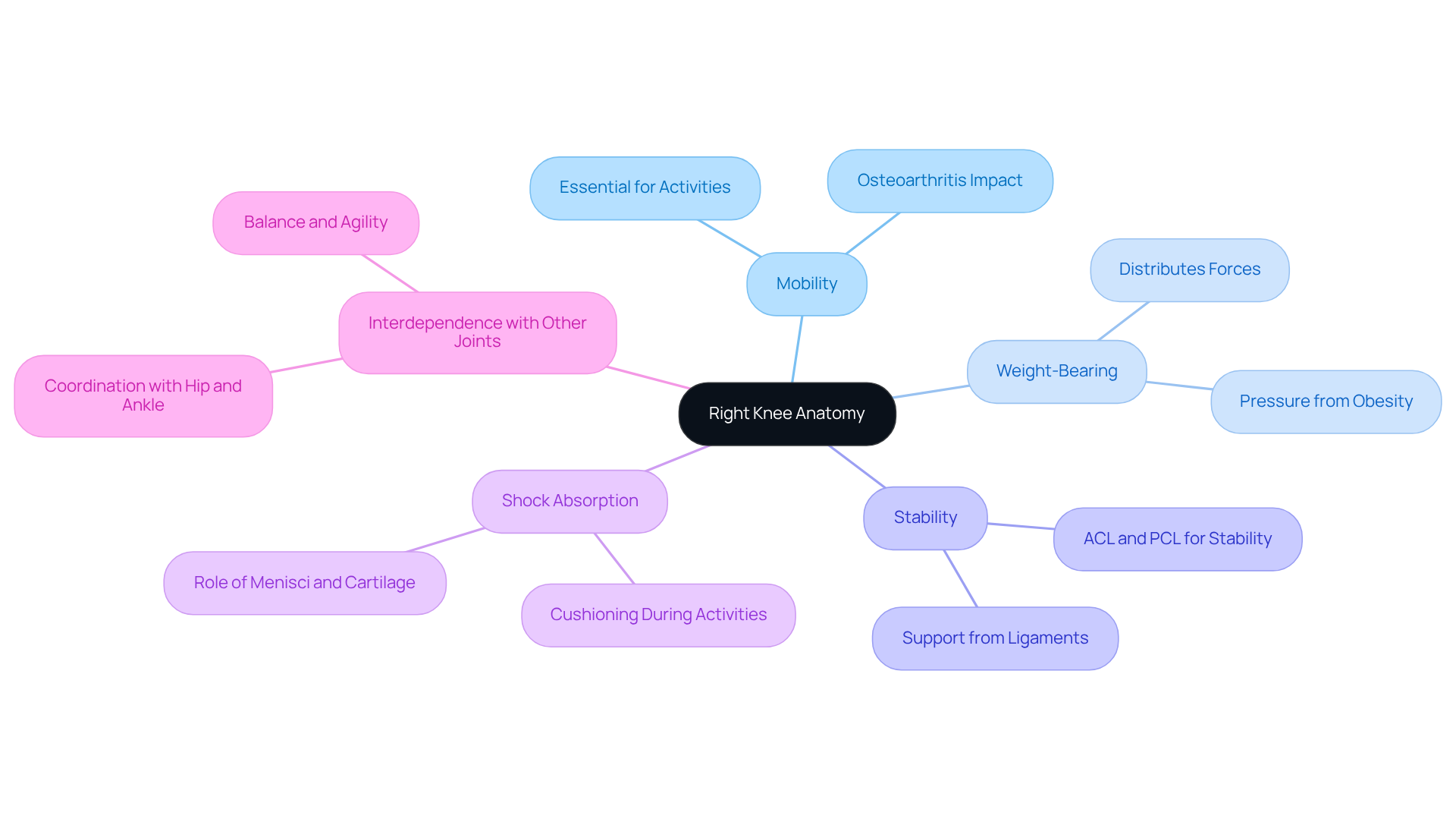
Many people struggle with knee pain that affects their daily lives and limits their ability to stay active. The knee joint is essential for mobility, allowing you to walk, run, jump, and climb stairs. This mobility is crucial for maintaining an active lifestyle.
The knee also bears your body weight during standing and movement, distributing forces to prevent harm. The menisci and cartilage absorb shock, protecting your bones from excessive stress. Unfortunately, osteoarthritis impacts over 32.5 million Americans, leading to pain, swelling, and stiffness that can significantly hinder your daily activities.
Stability is another key function of the knee joint. The ligaments surrounding it provide support, preventing excessive movement that could lead to injuries. The ACL and PCL are particularly important for keeping your knee stable during dynamic activities.
Additionally, the cartilage and menisci act as shock absorbers, cushioning your knee during high-impact activities. This function is vital for preventing wear and tear over time.
Your knee works in harmony with other joints, like the hip and ankle, to facilitate coordinated movement. This interdependence is crucial for activities that require balance and agility.
Understanding the right knee anatomy is crucial for anyone experiencing joint pain, as it can help guide you toward the appropriate care and support. Almost 25% of American adults deal with chronic knee discomfort, making it vital to understand how right knee anatomy affects your mobility and overall health. Injuries like meniscal tears and ligament damage can significantly hinder your mobility, highlighting the need for timely medical intervention and personalized treatment plans to restore function and reduce discomfort.
Evidence-based alternatives for pain management, such as exercise and strength training, can be particularly effective. Strengthening your quads and glutes can reduce pressure on the joint, while dietary changes, like adopting an anti-inflammatory diet, can further support joint health. Exploring minimally invasive solutions like Genicular Artery Embolization (GAE) may offer new avenues for treatment, but it’s essential to understand the associated risks for informed decision-making. Taking proactive steps now can help you regain control over your mobility and improve your quality of life.
Identify Common Injuries and Conditions Affecting the Right Knee
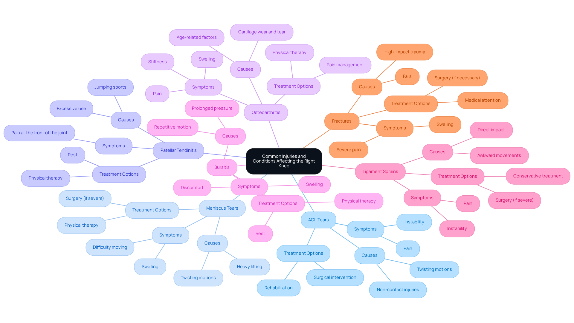
Living with joint pain can feel isolating and overwhelming, but you are not alone in this struggle. Common ailments and conditions affecting the right knee anatomy can significantly impact mobility and quality of life. Some of the most prevalent issues include:
- Anterior Cruciate Ligament (ACL) Tears: These injuries can cause instability and pain in the joint. It’s concerning to note that high school athletes, particularly females, are experiencing these injuries more frequently, with 57.5% of ACL injuries occurring without contact. Such injuries can disrupt your daily activities and lead to long-term challenges.
- Meniscus Tears: This discomfort can make even simple movements feel daunting. The menisci can be damaged due to twisting motions or heavy lifting, leading to swelling and difficulty moving the joint. Sudden meniscal tears often occur during sports activities involving pivoting, and treatment options vary based on the severity of the tear.
- Patellar Tendinitis: Also known as jumper’s knee, this condition arises from excessive use of the patellar tendon, leading to discomfort at the front of the joint. It is particularly common among athletes engaged in jumping sports, and it can be frustrating to deal with.
- Osteoarthritis: Osteoarthritis is a condition that affects the joints, causing the cartilage to wear down. This can lead to pain, stiffness, and swelling, making daily activities challenging. It’s prevalent among older adults and can significantly impair your quality of life.
- Bursitis: Inflammation of the bursae, small fluid-filled sacs that cushion the knee joint, can lead to discomfort and swelling. This often results from repetitive motion or prolonged pressure. Management typically includes rest and physical therapy, which can help ease your symptoms.
- Ligament Sprains: Sprains of the MCL or LCL can occur from direct impact or awkward movements, leading to pain and instability. These injuries are often treated conservatively unless severe, and it’s important to listen to your body during recovery.
- Fractures: Breaks around the joint, particularly of the patella or tibia, can result from falls or high-impact trauma, necessitating prompt medical attention. The incidence of knee fractures has been rising, highlighting the need for awareness and preventive measures.
Understanding the common injuries associated with right knee anatomy is crucial. With the right care, you can find relief and get back to doing what you love. Participation in ACL injury-prevention programs has been shown to effectively change landing mechanics, particularly in high-risk groups, emphasizing the importance of proactive steps today to pave the way for a more active and fulfilling tomorrow.
Conclusion
Many people struggle with knee pain that limits their daily activities. Understanding the anatomy of the right knee can be a key step toward finding relief and enhancing mobility. The knee joint, with its intricate structure of bones, ligaments, tendons, and cartilage, plays a pivotal role in facilitating movement and providing stability. Knowing how your knee works helps you spot problems early and find the right care for your situation.
Key components such as the femur, tibia, patella, and the surrounding ligaments and tendons work together to support daily activities and athletic endeavors. The functional importance of the knee cannot be overstated, as it bears body weight, absorbs shock, and maintains stability during movement. Common injuries, including ACL tears and osteoarthritis, highlight the importance of understanding your knee to get the help you need when it matters most. Innovative treatments, such as Genicular Artery Embolization, offer promising avenues for relief, underscoring the importance of seeking specialized care.
Ultimately, a proactive approach to knee health can lead to improved quality of life and enhanced mobility. By prioritizing education on knee anatomy and recognizing the signs of injury, individuals can take informed steps toward recovery. Engaging with healthcare professionals at Ponte Vedra Spine & Pain Center can provide the necessary support and guidance to navigate the complexities of knee health, ensuring you can reclaim your mobility and enjoy a more active life.
Frequently Asked Questions
What are the main components of the right knee anatomy?
The right knee joint consists of the patella (kneecap), femur (thigh bone), and tibia (shin bone). These bones work together, supported by ligaments, tendons, and cartilage.
How does the knee joint function?
The knee joint functions as a hinge, allowing for flexion and extension, while also permitting a limited range of rotation.
Why is understanding knee anatomy important for treating knee pain?
Understanding knee anatomy is crucial for addressing knee pain and finding appropriate treatment options, as each person’s experience with knee pain is unique.
What advancements have been made in joint anatomy research?
Recent advancements include the development of custom 3D-printed implants designed for individual joint anatomy, which have shown promise in improving surgical results and patient satisfaction.
How do personalized implants affect patient outcomes?
Case studies indicate that patients receiving personalized implants often report improved functionality and reduced recovery times, leading to better overall outcomes.
What is the commitment of Ponte Vedra Spine & Pain Center regarding knee treatment?
Ponte Vedra Spine & Pain Center is committed to using the latest technologies and personalized treatment plans to help patients overcome knee-related challenges and improve their quality of life.
List of Sources
- Define Right Knee Anatomy
- Advancements in knee replacement: More precise and personalized – Harvard Health
- Latest Advancements in Knee Surgery Recovery | Plancher Ortho
- Mayo Clinic Q&A: It’s a new era for knee replacements and other joint surgeries – Mayo Clinic News Network
- Can This “Living Knee” Revolutionize Joint Replacement?
- The latest knee replacement technology in 2025
- Explore Key Components of the Right Knee
- New Research Brings Joint Repair Closer for Millions With Osteoarthritis
- New biomaterial regrows damaged cartilage in joints
- New implant helps repair knee cartilage in UC Davis Health patients
- FDA OKs First in a New Generation of Knee Cartilage Repair
- Can This “Living Knee” Revolutionize Joint Replacement?
- Understand the Functional Importance of Right Knee Anatomy
- Identify Common Injuries and Conditions Affecting the Right Knee
Living with this?
We treat this in Ponte Vedra Beach.
Dr. Eli Loch, D.O. — double board-certified in anesthesiology and pain medicine. Reading about it only goes so far; a diagnosis tells you which of these actually applies to you.