
what is the root cause of sciatic nerve pain
What Is the Root Cause of Sciatic Nerve Pain?

Sciatica Describes a Pattern, Not One Diagnosis
Sciatica is a syndrome—a grouping of symptoms—rather than one specific injury or disease. The term describes a pattern of nerve-related pain, but it does not identify the structure causing that pain by itself.
Sciatica usually reflects irritation or compression of a lumbar or sacral nerve root that contributes to the sciatic nerve. In other words, the problem often begins near the lower spine before the nerve pathway continues through the buttock and leg. The sciatic nerve itself is not typically the structure being compressed.
That distinction matters because similar symptoms can arise from different sources. Disc tissue, bone changes, narrowing around the spinal canal, muscles near the buttock, or nearby joints can produce overlapping patterns. Your symptoms may suggest a location, but an examination is needed to determine whether the pattern fits a particular source.
A useful starting question is therefore not simply, “Where does the pain travel?” Ask which structure could be irritating a nerve, whether the physical findings support that possibility, and whether imaging—if needed—matches the clinical picture. This approach keeps a familiar symptom label from becoming an unsupported diagnosis.
The Usual Source Is a Lumbar or Sacral Nerve Root
Lumbar and sacral nerve-root irritation is the usual mechanism behind sciatica. The sciatic nerve is formed from nerve roots that leave the lower spine, and compression of one of those roots can create pain along the pathway supplied by that nerve.
The last lumbar nerve root, commonly identified as L5, and the first sacral nerve root, identified as S1, are often involved in the pattern called radiculopathy. Radiculopathy means that a nerve root is irritated or compressed near the spine. It does not mean that your symptoms have been confirmed as a particular spinal condition.
Several processes can place pressure on a nerve root. A disc may bulge or herniate, bone may narrow the available space, or other spinal changes may reduce room around the nerve. The location and degree of contact matter. A structure seen on an image is not automatically the source of pain.
The same broad symptom label can therefore describe different clinical situations. Two people may both report pain extending into a leg, yet one may have a disc-related nerve-root problem while the other has spinal narrowing or a nonspinal condition. The symptom pattern starts the evaluation; it does not finish it.
Nerve-root irritation can also produce symptoms beyond pain. Depending on the affected pathway, you may describe altered sensation or other changes during an examination. Those findings must be interpreted with the location, timing, and progression of your symptoms rather than treated as proof of one cause.
Common Spinal Causes Can Affect the Same Nerve Pathway
Spinal structures can irritate or compress the nerve roots that contribute to the sciatic nerve. Disc tissue and bone spurs are common sources of this pressure, although the same symptom pattern can arise from other processes affecting the lower spine.
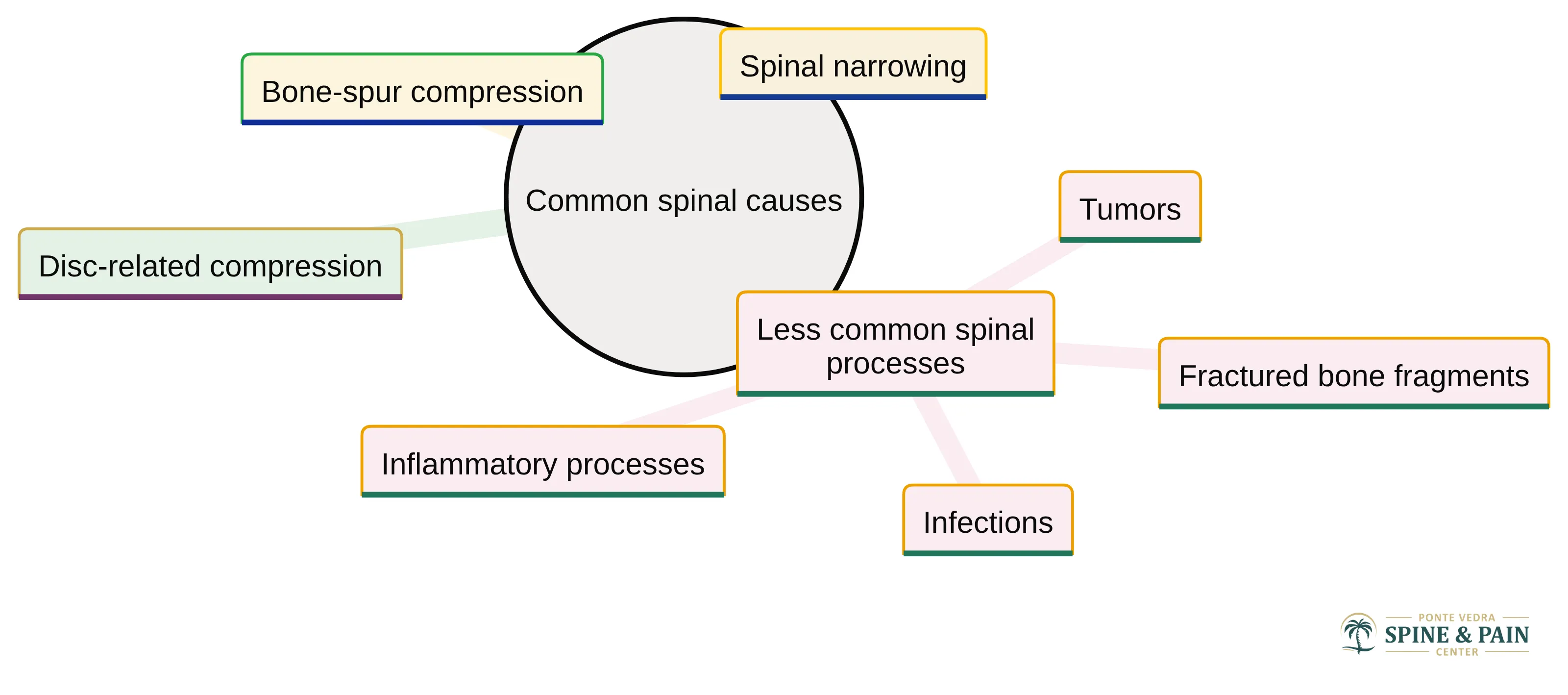
The main spinal possibilities include:
- Disc-related compression: Disc tissue can extend into space occupied by a nerve root and irritate it.
- Bone-spur compression: A bone spur can narrow the passage around a nerve root.
- Spinal narrowing: Changes that reduce room within or around the spinal canal can place pressure on nerves.
- Less common spinal processes: Fractured bone fragments, tumors, infections, or inflammatory processes can affect nerve roots.
These possibilities do not carry the same implications, and they cannot be separated by the word sciatica alone. The timing of symptoms, their distribution, findings on examination, and the presence or absence of other symptoms all help shape the evaluation.
A less common possibility still deserves appropriate consideration when the clinical picture does not fit an uncomplicated pattern. Persistent or unusual symptoms should be assessed rather than assigned to a familiar explanation without further evaluation. The purpose of that evaluation is to connect the suspected structure with the symptoms you actually experience.
Pain Distribution Can Suggest—but Cannot Prove—the Source
Pain distribution can suggest which nerve-root territory is involved, but it cannot prove the source of sciatic nerve pain. Sciatic pain often follows the skin territory supplied by an affected nerve root, known as a dermatome.
The pattern is not always neatly mapped onto the skin. Pain can also involve deeper tissues outside classic dermatomal areas, sometimes described as a dynatomal distribution. That broader pattern helps explain why pain location alone may not identify the precise structure involved.
You can note several useful details before an evaluation:
- Where the pain begins and where it travels
- Whether the pattern stays in the buttock or extends down the leg
- Whether sitting, standing, walking, or a particular position changes it
- Whether the pattern is stable or has changed over time
- Whether numbness, altered sensation, or weakness accompanies the pain
Those observations can make your history more useful. They cannot establish a diagnosis without the rest of the assessment, and a pattern that sounds typical does not exclude another source.
A Herniated Disc Matters Only When It Matches the Symptoms
A herniated disc can cause sciatica-like pain, but a herniated disc does not necessarily produce symptoms. Some herniated discs do not compress a nerve sufficiently to create noticeable pain or other nerve-related symptoms.
That distinction prevents a common interpretive error: treating the presence of a disc herniation as proof that it explains every symptom. The finding becomes more meaningful when its location corresponds with the side, distribution, and clinical findings associated with your pain.
A disc finding may therefore be:
- Relevant: It is located where it could affect the nerve associated with your symptoms, and the examination supports that connection.
- Incidental: It is visible on imaging but does not fit the symptoms or examination findings.
- Uncertain: The image and clinical picture do not align clearly, so further professional assessment is needed.
Imaging findings are most meaningful when they correspond with symptoms and clinical findings. A scan does not establish the pain source by itself, particularly because disc herniations can appear in people who have no symptoms or sciatica.
You should not treat the scan label as the diagnosis by itself. The useful question is whether the disc finding explains the whole clinical pattern, including the side of the pain, its distribution, its timing, and the examination findings.
Walking and Standing Patterns May Point Toward Spinal Narrowing
Spinal stenosis can narrow the spinal canal and place pressure on nerves, creating symptoms that resemble other forms of sciatica. The activity pattern may provide a useful clue, although it cannot confirm spinal narrowing by itself.
Symptoms associated with spinal stenosis may worsen with walking or standing and ease with sitting. That pattern differs from pain that changes primarily with another position or movement, but patterns can overlap. Your response to activity should therefore be recorded as part of the history rather than used as a self-diagnosis.
Pay attention to what happens during ordinary activities:
- Does standing bring on or intensify the symptoms?
- Does walking increase the leg pain?
- Does sitting ease the symptoms?
- Does the pattern remain consistent from day to day?
- Do symptoms limit how far you can walk or how long you can stand?
These details help a clinician compare possible sources. Spinal narrowing is one potential explanation among several, not a conclusion that can be made from walking tolerance alone. A careful evaluation can also consider whether the activity pattern reflects another spinal or extra-spinal source.
Not Every Sciatica-Like Symptom Begins in the Lower Back
Extra-spinal conditions can mimic sciatica even when the lumbar spine is not the starting point. Some symptoms arise along the sciatic nerve outside the spine, while others come from nearby muscles or joints and only resemble nerve-root pain.
Possible mimics include:
- Piriformis syndrome: Tightness or spasm in the piriformis muscle deep in the buttock can irritate or compress the sciatic nerve. Pain may remain localized to the buttock instead of radiating down the leg in a typical pattern.
- Hip or knee problems: A hip or knee disorder can refer pain into the leg without directly compressing the sciatic nerve.
- Other extra-spinal conditions: Quadratus lumborum myofascial pain syndrome, cluneal nerve disorder, and osteitis condensans ilii have also been described as possible sciatica mimics.
The distinction matters because treatment approaches differ when symptoms begin in the spine, outside the spine, or in a nearby joint. A lumbar-focused explanation may be incomplete if the pain pattern, examination, or activity response points elsewhere.
Pain that stays mainly in the buttock does not prove piriformis involvement, just as pain near the hip does not prove a joint disorder. These patterns can guide questions during an evaluation, but they do not replace examination findings.
You do not need to determine which mimic applies on your own. Bring the full pattern to an evaluation, including buttock pain, leg radiation, joint symptoms, and any activities that reproduce or ease the discomfort. The more clearly you describe those details, the easier it is to compare spinal and extra-spinal possibilities. The comparison does not always lead to a single source. Spinal and extra-spinal contributors can coexist, so identifying one plausible contributor does not automatically exclude another. A person may therefore have a nerve-root pattern alongside pain arising from the buttock, hip, knee, or another nearby structure. That overlap can make the symptom map seem inconsistent without establishing a particular diagnosis. It also explains why a prior label may not account for every feature of a new episode.
When describing symptoms, separate the components instead of compressing them into the word “sciatica.” Note where each sensation begins, whether it travels down the leg, which activities change it, and whether the components started together. Include what remains constant and what has changed. This organization gives a clinician a clearer basis for comparing spinal and extra-spinal possibilities, especially when no single pattern explains everything. The goal is not to identify the cause independently; it is to preserve enough detail for the evaluation to test whether one source, multiple contributors, or a mimic best fits the full picture.
Why an Examination Matters More Than a Scan Alone
Clinical evaluation begins with a careful medical history and physical examination. The history identifies the timing, distribution, triggers, and progression of symptoms, while the examination helps determine whether the findings fit a nerve-root pattern or suggest another source.
Imaging is often not needed at the initial assessment. A clinician may decide that the history and examination provide enough information to begin the evaluation without immediately ordering a scan. Imaging becomes more useful when the clinical situation calls for additional clarification or when symptoms and findings warrant a closer look.
This approach keeps the image in its proper role. A scan can show anatomy, but anatomy alone does not explain how that structure relates to your symptoms. The clinical question is not simply what appears on the image; it is whether the finding matches the side, distribution, examination, and course of the pain.
CT or MRI can show disc herniations in 20%–36% of people without symptoms or sciatica. That finding is one reason imaging should be interpreted alongside the clinical picture rather than treated as an independent explanation.
When an image and your symptoms do not correspond, the scan should not automatically determine the next step. When they do correspond, the match can help clarify which structure may be involved. A qualified clinician must interpret that relationship in context.
An evaluation can also identify when the familiar label no longer fits. A change in symptom distribution, a new functional limitation, or an unusual progression may alter which possibilities deserve attention. That is why a prior scan or previous explanation should not automatically be applied to every new episode.
Separate Familiar Radiating Pain from Warning Signs
New sciatica accompanied by bladder or bowel changes, urinary retention, or altered saddle sensation has been reported in acute cauda equina syndrome.
Seek prompt medical care if you develop:
- New difficulty passing urine or urinary retention
- New loss of bladder or bowel control
- New bowel dysfunction
- Altered or reduced sensation in the saddle region
Do not wait for a routine evaluation when these changes appear with new sciatica. This guidance is intentionally narrow: the listed symptoms require prompt assessment, while other pain patterns still need to be evaluated according to their severity, persistence, and progression.
If you are unsure whether a new change fits one of these warning signs, describe the symptom clearly when seeking care. Do not rely on a scan, an old diagnosis, or a familiar pain pattern to dismiss a new bladder, bowel, or saddle-sensation change.
How Source Identification Informs General Pain-Management Options
Source identification helps clinicians match pain-management options to the structure and mechanism under consideration. Spinal nerve-root pain, extra-spinal irritation, and a joint-related mimic should not automatically be approached as the same problem. The cited clinical review describes pain management as part of sciatica care and includes epidural treatments and radiofrequency among more interventional options.
The purpose of this distinction is practical. A treatment directed toward a spinal nerve root may not address pain referred from the hip, knee, or a muscle outside the spine. Conversely, a symptom pattern that suggests nerve-root irritation may require a different discussion from pain that remains localized near a joint or buttock.
These are general examples of image-guided treatments, not individualized instructions. Other general pain-management discussions may include nerve stimulation or radiofrequency ablation when those options are relevant to the clinical context. Their inclusion in a treatment discussion does not identify the pain source or establish that a particular option is appropriate. The evaluation remains the basis for relating any proposed intervention to the suspected structure and the patient’s findings. That relationship is why source identification comes before a procedure discussion.
The practical purpose of a targeted procedure is not to replace the evaluation.
You can use that distinction when discussing options: first clarify what source is being considered, then ask how the proposed intervention relates to that source. The discussion should connect your symptom pattern and examination findings with the treatment being considered, rather than relying on the procedure name alone.
Whether an option is appropriate depends on your history, examination, imaging when indicated, suspected source, and overall clinical circumstances. A treatment that makes sense for confirmed nerve-root irritation may not address pain referred from the hip, knee, or a muscle outside the spine.
Choose a Clear Next Step for Sciatic Nerve Pain in Ponte Vedra Beach
Persistent, recurrent, or concerning sciatic nerve pain deserves an evaluation that connects your symptoms with examination findings and, when appropriate, imaging. The central question is not only where the pain travels, but whether the source is a lumbar or sacral nerve root, a spinal structure, or an extra-spinal mimic.
If you are near Ponte Vedra Beach and need help clarifying the pattern, arrange a professional evaluation. Bring a concise description of when the pain began, where it travels, what changes it, and whether you have experienced any bladder, bowel, or saddle-sensation changes. We can discuss general pain-management and image-guided treatment options in the context of your evaluation; a qualified professional must determine individual suitability.
New sciatica with bladder or bowel changes, urinary retention, or altered saddle sensation requires prompt medical assessment rather than a routine appointment.
Questions to Ask Before Seeking Evaluation for Sciatic Nerve Pain
What are common causes of sciatic nerve pain?
Common causes include irritation or compression of lumbar or sacral nerve roots, often due to herniated discs, bone spurs, or spinal stenosis. Extra-spinal conditions like piriformis syndrome or hip issues can also mimic sciatica.
How can I distinguish between sciatica and other conditions?
Observing the pain’s distribution, triggers, and accompanying symptoms can help. Sciatica typically follows a nerve-root pattern, but other conditions may present similar symptoms without nerve involvement.
When should I seek medical evaluation for sciatic nerve pain?
Seek prompt medical evaluation if you experience new bladder or bowel changes, urinary retention, or altered sensation in the saddle area, as these may indicate serious conditions like cauda equina syndrome.
How can I prepare for a medical evaluation of my sciatic nerve pain?
Prepare by noting when the pain began, its location and distribution, any changes with activities, and any accompanying symptoms. This information will help your clinician assess your condition.
Living with this?
We treat this in Ponte Vedra Beach.
Dr. Eli Loch, D.O. — double board-certified in anesthesiology and pain medicine. Reading about it only goes so far; a diagnosis tells you which of these actually applies to you.